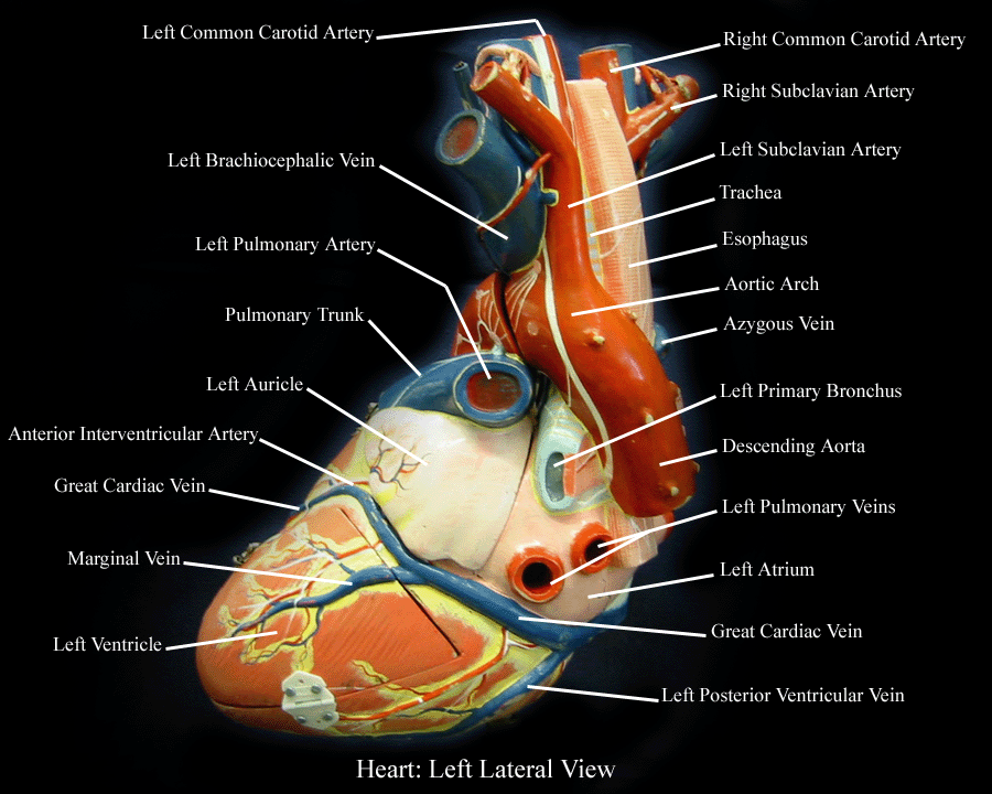
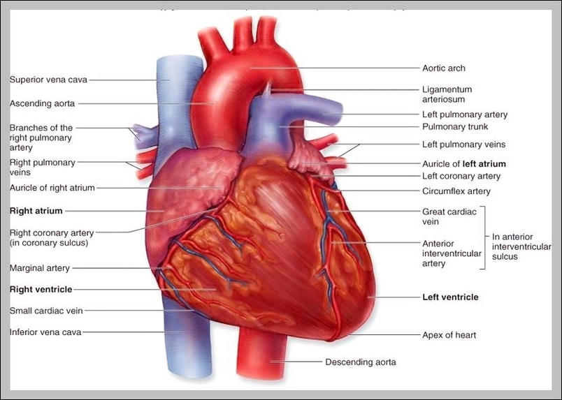
43 heart structure and labels
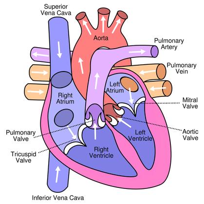
Structure of the Heart | The Franklin Institute The heart consists of four chambers: two atria on the top and two ventricles on the bottom. Looking at the Valentine's Day heart, the two rounded humps at the top are rounded like the top of a lower-case "a." The bottom is shaped like a "v." Feel it working What else is inside your heart? heart diagram and labels The Anatomy and Physiology of Animals/Circulatory System Worksheet. 11 Images about The Anatomy and Physiology of Animals/Circulatory System Worksheet : walls label label beginning Heart Diagram With Labels And Blood Flow, labelled diagram of heart a level - Clip Art Library and also labelled diagram of heart a level - Clip Art Library.
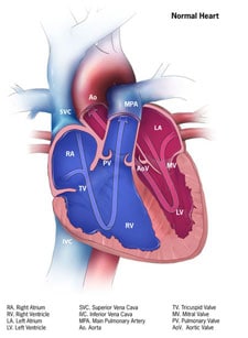
Human Heart (Anatomy): Diagram, Function, Chambers, Location in Body Human Heart (Anatomy): Diagram, Function, Chambers, Location in Body The right atrium receives blood from the veins and pumps it to the right ventricle. The right ventricle receives blood from the...

Heart structure and labels
Structure and Function of the Heart - Medical News The heart is the main organ in the circulatory system, the structure is primarily responsible for delivering blood circulation and transportation of nutrients in all parts of the body. This ... Heart Anatomy: Labeled Diagram, Structures, Function, and Blood Flow There are 4 chambers, labeled 1-4 on the diagram below. To help simplify things, we can convert the heart into a square. We will then divide that square into 4 different boxes which will represent the 4 chambers of the heart. The boxes are numbered to correlate with the labeled chambers on the cartoon diagram. Heart Labels - Printable or Custom Printed Stickers | Avery.com Use our free specialty shape label templates to easily personalize your heart labels online. Customize one of our free designs or upload your own graphics and then choose the printing option that works best for you. Order your blank heart labels or custom printed heart labels and stickers online and get free shipping on orders of $50 more.
Heart structure and labels. Diagrams, quizzes and worksheets of the heart | Kenhub Worksheet showing unlabelled heart diagrams. Using our unlabeled heart diagrams, you can challenge yourself to identify the individual parts of the heart as indicated by the arrows and fill-in-the-blank spaces. This exercise will help you to identify your weak spots, so you'll know which heart structures you need to spend more time studying ... Heart Anatomy | Anatomy and Physiology | | Course Hero Learning Objectives. By the end of this section, you will be able to: Describe the location and position of the heart within the body cavity. Describe the internal and external anatomy of the heart. Identify the tissue layers of the heart. Relate the structure of the heart to its function as a pump. Compare systemic circulation to pulmonary ... heart | Structure, Function, Diagram, Anatomy, & Facts | Britannica heart, organ that serves as a pump to circulate the blood. It may be a straight tube, as in spiders and annelid worms, or a somewhat more elaborate structure with one or more receiving chambers (atria) and a main pumping chamber (ventricle), as in mollusks. In fishes the heart is a folded tube, with three or four enlarged areas that correspond to the chambers in the mammalian heart. Human Heart Models | Heart Anatomy Models | Vitality Medical The heart model with labels is hand-painted with vivid colors to illustrate the papillary muscles, heart valves, and adjacent structures. Sort By 4 Items Magnetic Heart Model, Life Size, 5 Parts $327.45 View Details Human Heart Model $450.66 - $566.36 View Details Classic Heart Model $81.03 View Details Magnetic Heart Model, Life Size, 5 Part G01
Free Heart Worksheets for Human Anatomy Lessons Print out sheet of the human heart with labels - This fun heart worksheet shows kids the different parts of the heart. They'll learn about the left ventricle, the left atrium, the tricuspid valve, and more. Human Heart Clipart - There is a coloring page, heart labeling worksheet and heart anatomy chart. Clipart is a fun way for kids to ... Structure of the Heart | SEER Training Structure of the Heart. The human heart is a four-chambered muscular organ, shaped and sized roughly like a man's closed fist with two-thirds of the mass to the left of midline. The heart is enclosed in a pericardial sac that is lined with the parietal layers of a serous membrane. The visceral layer of the serous membrane forms the epicardium. Human Heart Diagram Labeled | Science Trends List Of Heart Structures Heart Chambers Ventricles - The bottom two heart chambers. Atra - The upper two heart chambers. Wall Of The Heart Sinoatrial Node - A collection of tissue that releases electrical impulses and defines the rate of contraction for the heart. Atrioventricular Bundle - The fibers which transmit cardiac impulses. How to Draw the Internal Structure of the Heart (with Pictures) To draw the internal structure of a human heart, follow the steps below. Part 1 Finding a Diagram 1 To find a good diagram, go to Google Images, and type in "The Internal Structure of the Human Heart". Find an image that displays the entire heart, and click on it to enlarge it. 2 Find a piece of paper and something to draw with.
Heart: Anatomy and Function - Cleveland Clinic What are the parts of the heart's anatomy? The parts of your heart are like the parts of a house. Your heart has: Walls. Chambers (rooms). Valves (doors). Blood vessels (plumbing). Electrical conduction system (electricity). Heart walls Your heart walls are the muscles that contract (squeeze) and relax to send blood throughout your body. Heart Diagram with Labels and Detailed Explanation The heart is located under the ribcage, between the lungs and above the diaphragm. It weighs about 10.5 ounces and is cone shaped in structure. It consists of the following parts: Heart Detailed Diagram Heart - Chambers There are four chambers of the heart . The upper two chambers are the auricles and the lower two are called ventricles. A Diagram of the Heart and Its Functioning Explained in Detail The heart blood flow diagram (flowchart) given below will help you to understand the pathway of blood through the heart.Initial five points denotes impure or deoxygenated blood and the last five points denotes pure or oxygenated blood. 1.Different Parts of the Body ↓ 2.Major Veins ↓ 3.Right Atrium ↓ 4.Right Ventricle ↓ 5.Pulmonary Artery ↓ 6.Lungs heart diagram and labels heart diagram and labels Cardiac Muscle Histology - Embryology we have 9 Images about Cardiac Muscle Histology - Embryology like Label the Heart Quiz, Anatomy of the Heart (Latin) and also Lab 2 Pig Heart-Labeled | Cardiovascular, Estudos. Read more: Cardiac Muscle Histology - Embryology embryology.med.unsw.edu.au
heart - a level biology student
The structure of the heart - Structure and function of the heart ... It is located in the middle of the chest and slightly towards the left. The heart is a large muscular pump and is divided into two halves - the right-hand side and the left-hand side. The...

Labeling the Heart
Heart Diagram with Labels and Detailed Explanation - BYJUS Diagram of Heart. The human heart is the most crucial organ of the human body. It pumps blood from the heart to different parts of the body and back to the heart. The most common heart attack symptoms or warning signs are chest pain, breathlessness, nausea, sweating etc. The diagram of heart is beneficial for Class 10 and 12 and is frequently ...

In this diagram they are showing the function of the heart as they have labels to the ...
Human Heart - Anatomy, Functions and Facts about Heart The human heart is divided into four chambers, namely two ventricles and two atria. The ventricles are the chambers that pump blood and atrium are the chambers that receive the blood. Among which, the right atrium and ventricle make up the "right portion of the heart", and the left atrium and ventricle make up the "left portion of the heart." 5.

Anterior View of Sheep Heart and Structures | Anatomy organs, Cardiology, Medical
The Anatomy of the Heart, Its Structures, and Functions The heart is the organ that helps supply blood and oxygen to all parts of the body. It is divided by a partition (or septum) into two halves. The halves are, in turn, divided into four chambers. The heart is situated within the chest cavity and surrounded by a fluid-filled sac called the pericardium. This amazing muscle produces electrical ...

Label Heart Structure | Medical Science Navigator
147 Heart Anatomy With Labels Premium High Res Photos Browse 147 heart anatomy with labels stock photos and images available, or start a new search to explore more stock photos and images. of 3. NEXT.
The Anatomy and Physiology of Animals/Circulatory System Worksheet - WikiEducator
Label the heart — Science Learning Hub Label the heart Interactive Add to collection In this interactive, you can label parts of the human heart. Drag and drop the text labels onto the boxes next to the diagram. Selecting or hovering over a box will highlight each area in the diagram. Right ventricle Right atrium Left atrium Pulmonary artery Left ventricle Pulmonary vein Semilunar valve

Pick Correct Options For Human Heart Quiz - ProProfs Quiz
Heart: illustrated anatomy - e-Anatomy - IMAIOS This interactive atlas of human heart anatomy is based on medical illustrations and cadaver photography. The user can show or hide the anatomical labels which provide a useful tool to create illustrations perfectly adapted for teaching. Anatomy of the heart: anatomical illustrations and structures, 3D model and photographs of dissection.
35 Label The Diagram Of The Heart - Labels Design Ideas 2020
Structure Of The Heart | A-Level Biology Revision Notes The heart is a hollow muscular organ that lies in the middle of the chest cavity. It is enclosed in the pericardium, which protects the heart and facilitates its pumping action. The heart is divided into four chambers: The two atria (auricles): these are the upper two chambers. They have thin walls which receive blood from veins.
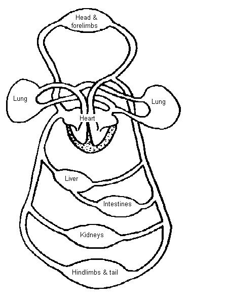
Dissection of the Frog
Layers of the heart: Epicardium, myocardium, endocardium - Kenhub The myocardium is functionally the main constituent of the heart and the thickest layer of all three heart layers. It is a muscle layer that enables heart contractions. Histologically, the myocardium is comprised of cardiomyocytes.Cardiomyocytes have a single nucleus in the center of the cell, which helps to distinguish them from skeletal muscle cells that have multiple nuclei dispersed in the ...

Fig 2 Gross Anatomy of Heart (a) Quiz
Heart Blood Flow | Simple Anatomy Diagram, Cardiac Circulation ... - EZmed One of the first things you will notice if you look at the 12 steps is the pattern between the right and left side of the heart is similar. Step 1 and 6 involve a blood vessel, which makes sense as this is how blood enters and exits that side of the heart. Steps 2-5 involve a chamber, valve, chamber, and valve.

Cuthbert - 7th Grade Science Day to Day: Comparing Plant and Animal Cells
Label Heart Anatomy Diagram Printout - EnchantedLearning.com Every day, the heart pumps about 2,000 gallons (7,600 liters) of blood, beating about 100,000 times. Label the heart anatomy diagram below using the heart glossary. Note: On the diagram, the right side of the heart appears on the left side of the picture (and vice versa) because you are looking at the heart from the front. Enchanted Learning Search

The Heart | S-cool, the revision website
Heart Labels - Printable or Custom Printed Stickers | Avery.com Use our free specialty shape label templates to easily personalize your heart labels online. Customize one of our free designs or upload your own graphics and then choose the printing option that works best for you. Order your blank heart labels or custom printed heart labels and stickers online and get free shipping on orders of $50 more.
12+ Model Heart Labeled | Robhosking Diagram
Heart Anatomy: Labeled Diagram, Structures, Function, and Blood Flow There are 4 chambers, labeled 1-4 on the diagram below. To help simplify things, we can convert the heart into a square. We will then divide that square into 4 different boxes which will represent the 4 chambers of the heart. The boxes are numbered to correlate with the labeled chambers on the cartoon diagram.

Heart Anatomy Part 1 - YouTube
Structure and Function of the Heart - Medical News The heart is the main organ in the circulatory system, the structure is primarily responsible for delivering blood circulation and transportation of nutrients in all parts of the body. This ...
Congenital Heart Defects - How the Heart Works | CDC
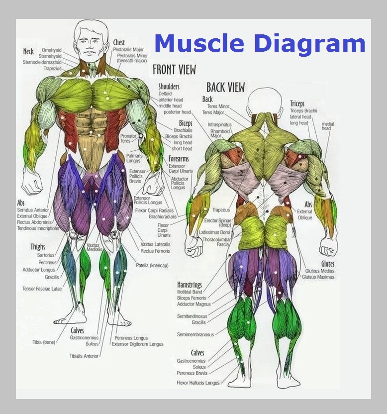
Muscle Diagram – Graph Diagram
32 Label The Diagram Of The Heart - Label Design Ideas 2020
Post a Comment for "43 heart structure and labels"